- Anaplastic astrocytoma
A World Health Organization (WHO) grade III tumor that originates from astrocytes.1
Histologic features include one or more of the following2:
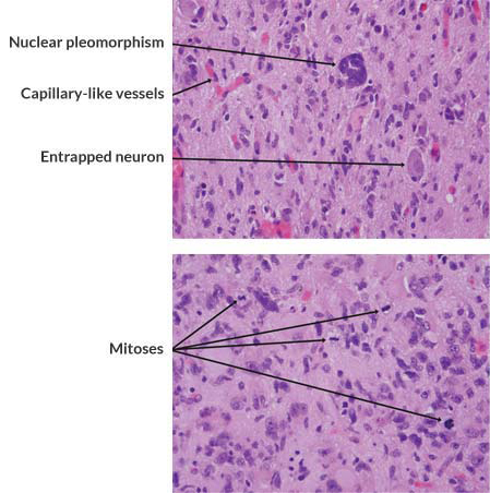
- Elevated mitotic activity (>1 mitosis identified throughout)—most defining criterion
- Increased cellularity
- Nuclear pleomorphism (distinct atypical nuclei)
Morphology (cell shape, cytoplasmic appearance, and shape of nuclei) provides the means for distinguishing astrocytic (irregular nuclear contour, angulation) from oligodendroglial tumors (regular nuclear contour, round to oval), which have more regular nuclei.2,3

Astrocytic cells are characterized by angulated hyperchromatic nuclei. Note the elevated mitotic activity.5
Micrographs courtesy of Eyas M. Hattab, MD. Indiana University School of Medicine.
WHO grade III gliomas are defined as having mitotic activity, clear infiltrative capabilities, and anaplasia.4
REFERENCES
- American Brain Tumor Association. About Brain Tumors: A Primer for Patients and Caregivers, Chicago, IL: American Brain Tumor Association; 2018.
- Brat DJ, Prayson RA, Ryken TC, Olson JJ. Diagnosis of malignant glioma: role of neuropathology. J Neurooncol. 2008;89:287-311.
- Perry A, Aldape KD, George DH, Burger PC. Small cell astrocytoma: an aggressive variant that is clinicopathologically and genetically distinct from anaplastic oligodendroglioma. Cancer. 2004;101:2318-2326.
- Huttner A. Molecular neuropathology and the ontogeny of malignant gliomas. In: Gunel JM, Piepmeir JM, Baehring JM, eds. Malignant Brain Tumors. Cham, SUI: Springer International Publishing; 2017:18-19.
- Brandes AA, Nicolardi L, Tosoni A, et al. Survival following adjuvant PCV or temozolomide for anaplastic astrocytoma. Neuro Oncol. 2006;8:253-260.
- Anaplastic
oligodendrogliomaa World Health Organization (WHO) grade IIIa tumor that originates from oligodendrocytes.1
Histologic features include1,2:
- Perinuclear cytoplasmic clearing, also known as fried egg appearance (formalin fixation artifact; not seen on frozen section preps or smears)
- Microcalcifications
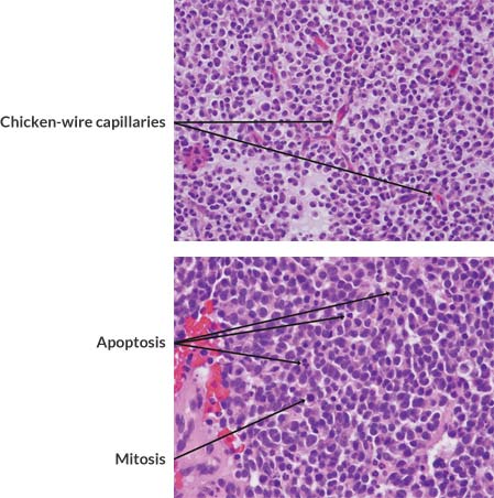
- Delicate branching capillaries (chicken-wire vasculature)
- Microcysts
This tumor is characterized by round, regular, monotonous nuclei, with little variability seen among cells.2
The 2007 WHO classification does not recognize a grade IV oligodendroglioma and hence the presence of vascular proliferation and/or necrosis does not qualify an otherwise anaplastic oligodendroglioma for the diagnosis of glioblastoma.4

Cellular uniformity is a consistent feature of oligodendroglioma, including anaplastic examples. Branching capillary-like blood vessels (chicken-wire vasculature) are characteristic.3
Micrographs courtesy of Eyas M. Hattab, MD. Indiana University School of Medicine.
REFERENCES
- American Brain Tumor Association. About Brain Tumors: A Primer for Patients and Caregivers, Chicago, IL: American Brain Tumor Association; 2018.
- Brat DJ, Prayson RA, Ryken TC, Olson JJ. Diagnosis of malignant glioma: role of neuropathology. J Neurooncol. 2008;89:287-311.
- Perry A, Aldape KD, George DH, Burger PC. Small cell astrocytoma: an aggressive variant that is clinicopathologically and genetically distinct from anaplastic oligodendroglioma. Cancer. 2004;101:2318-2326.
- Louis DN, Ohgaki H, Wiestler OD, et al. The 2007 WHO classification of tumours of the central nervous system. Acta Neuropathol. 2007;114:97-109.
- Anaplastic
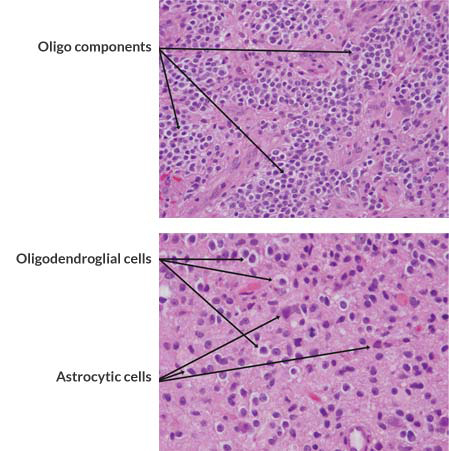
oligoastrocytomaa World Health Organization (WHO) grade III tumor that contains both oligodendroglial cells and astrocytes1
Specific characteristics include1:
- Nuclear atypia/pleomorphism
- High cellularity
- High mitotic activity
- Microvascular proliferation
The WHO classification suggests that features of anaplasia should be present. Anaplasia may be observed in oligodendroglial cells, astrocytes, or in both components of the tumor.1
Anaplastic oligoastrocytomas with necrosis should be classified as glioblastoma with oligodendroglial component. Reporting of the oligodendroglial component is important as such tumors may carry a better prognosis than standard glioblastoma. The finding of necrosis has been associated with significantly decreased survival.1

Most oligoastrocytomas show a mixture of tumor cells with oligodendroglial and astrocytic features.1
Micrographs courtesy of Eyas M. Hattab, MD. Indiana University School of Medicine.
REFERENCES
- Brat DJ, Prayson RA, Ryken TC, Olson JJ. Diagnosis of malignant glioma: role of neuropathology. J Neurooncol. 2008;89:287-311.
- Glioblastoma
a WHO grade IV tumor.2
Specific characteristics include1:
- Increased cellularity
- Distinct nuclear atypia
- High degree of mitotic activity
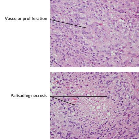
- Vascular proliferation
- Necrosis (geographic or palisading)
WHO grade IV lesions are generally associated with a rapid preoperative and postoperative progression and fatal outcomes2
Glioblastoma was reported as the most commonly occurring malignant brain and other CNS tumor (14.6% of all tumors and 48.3% of malignant tumors).3

The identification of vascular proliferation permits the diagnosis of glioblastoma in diffuse astrocytic neoplasms.1
Micrographs courtesy of Eyas M. Hattab, MD. Indiana University School of Medicine.
REFERENCES
- Brat DJ, Prayson RA, Ryken TC, Olson JJ. Diagnosis of malignant glioma: role of neuropathology. J Neurooncol. 2008;89:287-311.
- National Cancer Institute. Adult Brain Tumors (PDQ®). 2020. Available at: https://www.cancer.gov/types/brain/hp/adult-brain-treatment-pdq#_5 Accessed April 2020.
- Ostrom QT, Cioffi G, Gittleman H, Patil N, Waite K, Kruchko C, Barnholtz-Sloan JS. CBTRUS Statistical Report: Primary Brain and Other Central Nervous System Tumors Diagnosed in the United States in 2012-2016. Neuro Oncol. 2019 Nov 1;21(Supplement_5):v1-v100.
High-Grade Gliomas
Select the tumor type below to learn more about its pathology
Important Safety Information | The following Warnings and Precautions have been associated with the use of GLIADEL Wafer: seizures, intracranial hypertension, impaired neurosurgical wound healing, meningitis, and wafer migration. GLIADEL Wafer can cause fetal harm when administered to a pregnant woman.
Please see Important Safety Information, as well as the Full Prescribing Information.
INDICATIONS
GLIADEL Wafer is indicated for the treatment of patients with newly-diagnosed high-grade glioma as an adjunct to surgery and radiation.
GLIADEL Wafer is also indicated for the treatment of patients with recurrent glioblastoma as an adjunct to surgery.
IMPORTANT SAFETY INFORMATION
WARNINGS AND PRECAUTIONS
Seizures: Seizures occurred in 37% of patients treated with GLIADEL Wafers for recurrent glioma in the recurrent high-grade glioma trial. New or worsening (treatment emergent) seizures occurred in 20% of patients; 54% of treatment emergent seizures occurred within the first 5 post-operative days. The median time to onset of the first new or worsened post-operative seizure was four days. Institute optimal anti-seizure therapy prior to surgery. Monitor patients for seizures postoperatively.
Intracranial Hypertension: Brain edema occurred in 23% of patients with newly diagnosed glioma treated with GLIADEL Wafers in the newly-diagnosed high-grade glioma trial. Additionally, one GLIADEL-treated patient experienced intracerebral mass effect unresponsive to corticosteroids which led to brain herniation. Monitor patients closely for intracranial hypertension related to brain edema, inflammation, or necrosis of the brain tissue surrounding the resection. In refractory cases, consider re-operation and removal of GLIADEL Wafers or Wafer remnants.
Impaired Neurosurgical Wound Healing: Impaired neurosurgical wound healing including wound dehiscence, delayed wound healing, and subdural, subgaleal, or wound effusions occur with GLIADEL Wafer treatment. In the newly-diagnosed high-grade glioma trial, 16% of GLIADEL Wafer-treated patients with newly diagnosed glioma experienced impaired intracranial wound healing and 5% had cerebrospinal fluid leaks. In the recurrent high-grade glioma trial, 14% of GLIADEL Wafer-treated patients with recurrent high-grade glioma experienced wound healing abnormalities. Monitor patients post-operatively for impaired neurosurgical wound healing.
Meningitis: Meningitis occurred in 4% of patients with recurrent glioma receiving GLIADEL Wafers in the recurrent high-grade glioma trial. Two cases of meningitis were bacterial; one patient required removal of the Wafers four days after implantation; the other developed meningitis following reoperation for recurrent tumor. One case was diagnosed as chemical meningitis and resolved following steroid treatment. In one case the cause was unspecified, but meningitis resolved following antibiotic treatment. Monitor postoperatively for signs of meningitis and central nervous system infection.
Wafer Migration: GLIADEL Wafer migration can occur. To reduce the risk of obstructive hydrocephalus due to wafer migration into the ventricular system, close any communication larger than the diameter of a Wafer between the surgical resection cavity and the ventricular system prior to Wafer implantation. Monitor patients for signs of obstructive hydrocephalus.
Embryo-Fetal Toxicity: GLIADEL Wafers can cause fetal harm when administered to a pregnant woman. Carmustine, the active component of GLIADEL Wafer, is embryotoxic and teratogenic in rats at exposures less than the exposure at the recommended human dose based on body surface area (BSA) and embryotoxic in rabbits at exposures similar to the exposure at the recommended human dose based on BSA. Advise patients of the potential risk to a fetus. Advise females of reproductive potential to use effective contraception for 6 months after implantation of GLIADEL Wafer. Advise males with female partners of reproductive potential to use effective contraception for 3 months following implantation of Gliadel Wafers.
ADVERSE REACTIONS
The most common adverse reactions in newly-diagnosed high-grade glioma patients (incidence >10% and between arm difference ≥4%) are cerebral edema, asthenia, nausea, vomiting, constipation, wound healing abnormalities and depression.
The most common adverse reactions in recurrent high-grade glioma patients (incidence >10% and between arm difference ≥4%) are urinary tract infection, wound healing abnormalities and fever.
The Important Safety Information does not include all the information needed to use GLIADEL safely and effectively. For additional safety information, please consult the full Prescribing Information for GLIADEL.
To report SUSPECTED ADVERSE REACTIONS, contact Azurity Pharmaceuticals, Inc. at 1-800-461-7449 or FDA at www.fda.gov/medwatch or call 1-800-FDA-1088.

